Current Research
The goal of the Collins Lab is to integrate non-invasive imaging methods with experimental mechanics and computational modeling in order to develop clinically applicable tools to monitor bone integrity, fracture risk, and fracture healing in patients. Through this, the lab aims to better understand and model severe fracture types and cases with poor clinical outcomes to improve injury countermeasures and treatment strategies.
Assessing the Effects of Histotripsy on Osteosarcoma Tumor-Affected Bone
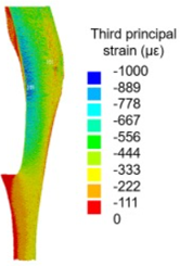
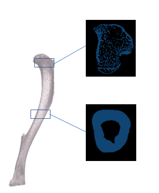
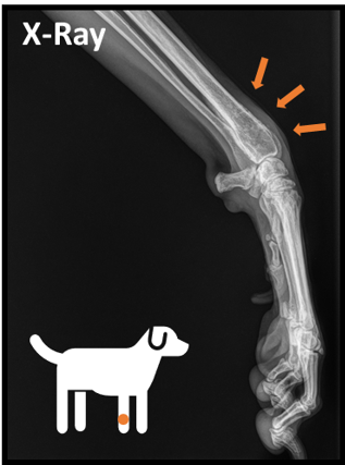
Evaluate Crash Factors Impacting Distal Tibia Fracture Severity using the Fracture Liberation Energy
Motivation: Distal tibia fractures are associated with high rates of clinical complications and frequently result in long-term functional limitations. Current measures of injury severity do not account for pathomechanical factors which contribute to persisting issues for clinical treatment and clinical prognosis (Work of Garrett Bangert).
Goal: The Fracture Liberation Energy is a quantitative severity score which relates the surface area created by a fracture to the energy dispersed in the distal tibia. The goal of this study is to characterize the relationship between vehicle crashworthiness, occupant demographics, crash mechanics and pathmechanical factors of injury severity.
The image above shows the process of identifying surface area liberated by a fracture (de novo surface) to compute the Fracture Liberation Energy. Please reference the work Dr. Kevin Dibbern, Dr. Don Anderson, Dr. Thomas Thaddeus and Dr. Christina Beardsley.
Analyzing the Morphology of Successful Anterior Cervical Discectomy and Surgical Site Fusions using X-Ray Imaging
Motivation: Spinal fusions are performed to treat degenerative disks and stabilize the spine. Current methods to determine the success of fusion require various radiology imaging techniques, are subjective largely depending on reviewer expertise, and can require invasive surgery. (Work of Alejandro Venable-Croft)
Goal: Develop a novel image-based analysis of successful spinal fusion using a Hurst/Variable Orientation Transform to detect sufficient bone growth into the fusion region.

Hurst/Variable Orientation Transform (H/VOT) is a fractal geometry technique that can characterize the surface topography of spinal fusions over time. H/VOT samples pixel values in a specified region of interest to compute the maximum difference in pixel intensity in all directions.
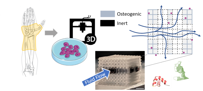
Exploring Breast Cancer Cell Recruitment and Survival in the Bone Niche using 3D-Printed Tissue-Engineered Bone Models
Motivation: With very high failure rates of anti-cancer drugs and deficiency to mimic physiological bone environment, it is clear that current gold standard (2D culture and animal models) lack in information to accurately predict effects of drugs and described the interactions in a multicell environment. (Work of Edward Shangin)
Goal: To understand the underlying parameters that influence breast-to-bone metastasis via a 3D co-culture tissue model that assesses the role of extracellular vesicles has on breast cancer development.

Research Partners